
Optical Spectroscopy
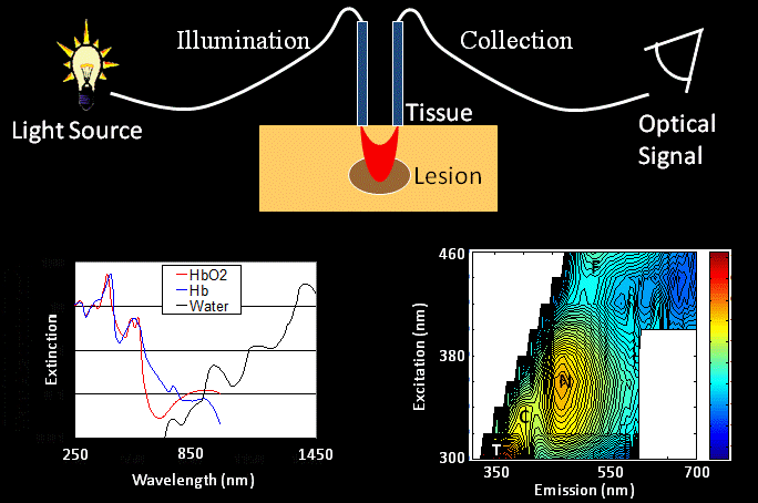
Optical spectra can be collected over a tissue volume with optical fibers that deliver light to the tissue surface and collect the diffusely back-scattered light. This approach allows for rapid interrogation of macroscopic volumes of tissue. The main absorbers in tissue in the visible and near infrared wavelength range include oxygenated and de-oxygenated hemoglobin and water. The main fluorophores in tissue that emit in the visible wavelength range include tryptophan (T, an amino acid), collagen (C, a structural protein) and the metabolic co-enzymes NADH (N) and FAD (F). Thus, optical spectroscopy of tissue allows for quantification of blood content and oxygenation, protein content and metabolic status. These tools are more practical for clinical applications than high-resolution optical imaging, and thus provide a method for translating pre-clinical imaging results to the clinic.
References
MC Skala, GM Palmer, KM Vrotsos, A Gendron-Fitzpatrick, N Ramanujam. Comparison of a Monte Carlo based physical model and principal component analysis for the diagnosis of normal and neoplastic epithelial tissues in vivo using diffuse reflectance spectroscopy. Optics Express, (15):12; 7863-7875 (2007).
MC Skala, GM Palmer, C Zhu, Q Liu, KM Vrotsos, CL Marshek-Stone, A Gendron-Fitzpatrick and N Ramanujam. Investigation of fiber-optic probe designs for optical spectroscopic diagnosis of epithelial pre-cancers. Lasers in Surgery and Medicine 34; 25-38 (2004).