
Optical Coherence Tomography
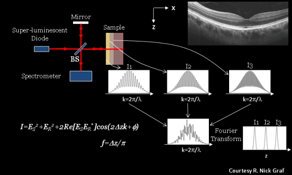
Optical Coherence Tomography (OCT) is a three-dimensional tissue imaging technique. Similar to ultrasound, OCT provides cross-sectional images of tissue, but does not require coupling media and provides cellular resolution (~1 to 10 micron isotropic resolution). The principles of OCT are based on the Michelson interferometer (above). Path length differences between the reference arm (mirror) and scatterers within the sample (such as cellular organelles) are detected at the spectrometer. The intensity of the reflections at different depths within the sample is determined from the detected interferogram (represented by the cosine term in the intensity (I) equation). Reflections at different depths in the sample
result in different frequency components in the detected interferogram. These depth-dependent signals within the interferogram can be deconvolved with the Fourier Transform to provide intensity as a function of depth in the sample. The sample is raster-scanned in two dimensions (x- and y-), and the interferogram provides depth-dependent information, resulting in a three-dimensional image of the tissue. The example OCT image shown here is of the human retina.
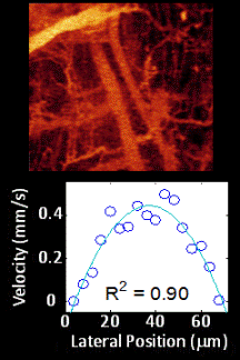
OCT is highly sensitive to movement in the sample (i.e. path length differences between the reference and sample arms) through changes in the phase of the Fourier Transformed data or through changes in the speckle pattern of the intensity image.
References
MC Skala, A Fontanella, L Lan, JA Izatt, and MW Dewhirst. Longitudinal optical imaging of tumor metabolism and hemodynamics Journal of Biomedical Optics 15:(1); 011112 (2010).
MC Skala, A Fontanella, H Hendargo, MW Dewhirst, and JA Izatt. Combined Hyperspectral and Optical Coherence Tomography Microscope for Noninvasive Hemodynamic Imaging. Optics Letters 34:(3); 289-291 (2009).