
Fluorescence Lifetime
When a fluorophore is excited, the emitted photon is released at a later (characteristic) time called the lifetime (tau). The fluorescence lifetime of organic fluorophores is usually on the order of picoseconds (10-12 seconds) to nanoseconds (10-9 seconds). The fluorescence lifetime is highly sensitive to the fluorophore microenvironment, including local changes in pH, O2, temperature and molecular binding. The metabolic co-enzymes NADH and FAD, which are found intrinsically in cells, have a double-exponential fluorescence lifetime decay attributed to their free and protein-bound states. Changes in the fluorescence lifetimes of these molecules have been detected with hypoxia and with early cancer development. These time-resolved kinetics can be used to diagnose cancer and monitor metabolic changes associated with cancer therapy. Fluorescence lifetime imaging is advantageous over simple intensity-based imaging because it is self-referenced and thus does not require careful calibration with a standard. It is also capable of resolving molecular species that cannot be differentiated with intensity- or wavelength-resolved methods.
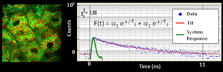
In fluorescence lifetime imaging, the color-coded value of each pixel in an image represents the mean fluorescence lifetime of all the fluorophores in that pixel. The fluorescence lifetime is determined by counting all the photons that arrive at the detector into discrete time bins. After detecting many photons, a decay curve emerges that is fit to a multi-exponential decay (after accounting for the finite time response of the system). The decay constants of this fit (tau) are the lifetime components of the pixel of interest.
References
MC Skala, KM Riching, A Gendron-Fitzpatrick, KW Eliceiri, and N Ramanujam. In vivo multiphoton microscopy of metabolic oxidation-reduction states and fluorescence lifetimes in normal and pre-cancerous epithelia. Proceedings of the National Academy of Sciences USA 104:(49); 19494-19499, (2007).
MC Skala, KM Riching, DK Bird, A Gendron-Fitzpatrick, KW Eliceiri, PJ Keely, and N Ramanujam. In vivo multiphoton fluorescence lifetime imaging of free and protein-bound NADH in normal and pre-cancerous epithelia. Journal of Biomedical Optics, 12:(2); 024014 (2007).