
RS-OCT For Imaging the Skin
The current standard for diagnosis of suspicious skin lesions involves biopsy followed by histopathology. In patients with multiple questionable lesions, clinicians are presented with the challenge of deciding which and how many skin lesions to biopsy, usually relying on visual inspection and palpation. Although this protocol for skin lesion diagnosis is presently the gold-standard, it is also subjective, invasive, time-consuming, and costly. These limitations have motivated the development of non-invasive techniques capable of direct characterization of questionable skin lesions. Such tools have the potential to assist in biopsy guidance, or possibly even circumvent the need for histology all together.
Optical techniques have demonstrated the potential to perform non-invasive characterization of skin lesions. Imaging modalities such as optical coherence tomography (OCT) are advantageous because they allow real-time, non-invasive visualization of tissue structure with micron-scale resolution. OCT is capable of visualizing the subsurface features of skin lesions, however achieving a definitive diagnosis based simply on maps of tissue reflectivity without much knowledge of the biochemical composition of the lesion can be difficult. Spectroscopic techniques such as Raman spectroscopy (RS) have demonstrated the ability to perform disease classification based on the molecular composition of skin lesions. Unfortunately, the inherently weak nature of Raman scattering makes it difficult to design a fast, low-cost Raman-imaging instrument for clinical applications. To date, clinical implementations of RS have been limited to point-wise measurements, which are susceptible to sampling error and have a limited ability to determine the spatial features of disease. In summary, RS excels at characterizing the biochemical composition of tissue with limited spatial information, while OCT excels at imaging tissue microstructure with limited biochemical specificity. Thus, the uniquely complementary strengths and limitations of RS and OCT make the two modalities well suited for combination into a single instrument.
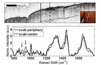 In Vivo RS-OCT of a scab on the back of a finger. In the OCT image (top), the center of the scab is clearly seen as ahyperreflective (dark) region in the center of the image. The vertical lines in the OCT image depict the axis from which Raman spectra were acquired. The Raman spectrum from the center of the scab exhibits subtle differences (arrows) in spectral regions corresponding to peaks seen in collagen and keratin
Our group has previously reported the development of the first combined RS-OCT instrument in a benchtop configuration and demonstrated its potential for performing in-vivo measurements in the skin. OCT complements the clinical implementation of RS by guiding the axis of spectral acquisition in order to reduce the likelihood of sampling error. RS can likewise complement the clinical application of OCT by performing highly specific biochemical characterization of tissues with irregular microstructural features. We have since miniaturized the benchtop RS-OCT instrument into a probe capable of performing clinical measurements of suspicious skin lesions. We are currently investigating the potential of this device to perform non-invasive characterization of potentially cancerous skin lesions.
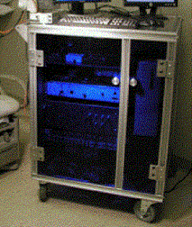
The combined RS-OCT clinical system
References
H. Krishnamoorthi, C. A. Patil, S. Munoz, G. Gutierrez, and A. Mahadevan-Jansen, “Non-destructive measurement of calvaria thickniss with Optical Coherence Tomography,” In preparation.
C. A. Patil, H. Krishnamoorthi, S. Munoz, G. Gutierrez, and A. Mahadevan-Jansen, “Rapid Measurement of Calvaria Volume and Biochemical Properties with Combined Raman Spectroscopy-Optical Coherence Tomography,” In preparation.
H. Krishnamoorthi, C.A. Patil, D.S. Perrien, E.C. O’Quinn, G. Gutierrez, J.S. Nyman, and A. Mahadevan-Jansen. “Optical analysis of physical and biochemical properties of murine calvaria with combined Raman spectroscopy optical coherence tomography.” Paper 7548F-149 presented at SPIE Photonics West, San Francisco, California, January 23-28, 2010.
C.A. Patil, J. Kalkman, D.J. Faber, J.S. Penn, T.G. van Leeuwen, and A. Mahadevan-Jansen “Structural and biochemical characterization of the rat retina with combined Raman spectroscopy optical coherence tomography.” Paper 7550-47 presented at SPIE Photonics West, San Francisco, California, January 23-28, 2010.
C. A. Patil, N. Bosschaart, M. D. Keller, T. G. van Leeuwen, and A. Mahadevan-Jansen, "Combined Raman spectroscopy and opti l coherence tomography device for tissue characterization," Optics Letters, 33 (10), 1135-1137 (2008)http://www.opticsinfobase.org/abstract.cfm?URI=ol-33-10-1135