
Analysis of Breast Tumor Margins with Raman Spectroscopy
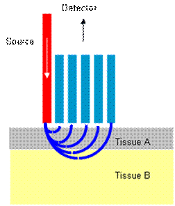
The treatment of breast tumors often involves breast-conserving therapy (BCT), in which the tumor is removed with a surrounding layer of healthy tissue known as the margin. In order to ensure that a large enough layer of healthy tissue surrounding the tumor has been removed, current methods require about a week of expensive analysis, often involving another surgery. Raman spectroscopy has shown the ability to distinguish between malignant and healthy tissue, particularly in the breast. Raman spectroscopy can be used to gain depth resolved information by creating a spatial offset between the laser source and detectors. This research uses the depth resolved Raman information to evaluate the completeness of tumor removal in the operating room.
References
X. Bi, B. Rexer, C. L. Arteaga, M. Guo, M. Li, A. Mahadevan-Jansen, Determination of HER2 amplification status in breast cancer cells using Raman spectroscopy, 2010, Proc. SPIE, Vol. 7560, 7560-30 (2010).
Keller, MD, Vargis E, Granja N, Meszoely I, Kelley M, A Mahadevan-Jansen. In vitro and in vivo SORS measurements for breast tumor surgical margin analysis. SPIE: Photonics West 2010. San Francisco, CA, January 2010.
Keller MD, Majumder SK, Kelley M, Mahadevan-Jansen A, Depth-resolved measurements in breast tissues with spatially offset Raman spectroscopy, In: Advanced Biomedical and Clinical Diagnostic Systems VII, BiOS 2009, Photonics West, San Jose CA, January 2009.
Keller MD, Majumder SK, Kelley MC, Meszoely I, Boulos FI, Olivares GM, and Mahadevan-Jansen A, Optical guidance in breast concerving therapy. In: Advanced Clinical Diagnostics Systems, BiOS 2008, San Jose, CA.
Keller MD and Mahadevan-Jansen A. Spatially Offset Raman Spectroscopy for Depth-sensitive Measurements in Excised Breast Tissues, FACSS 2008 (Federation of Analytical Chemistry and Spectroscopy Societies), September 2008.