Vanderbilt "Day of Light" Symposium

Abstracts
|
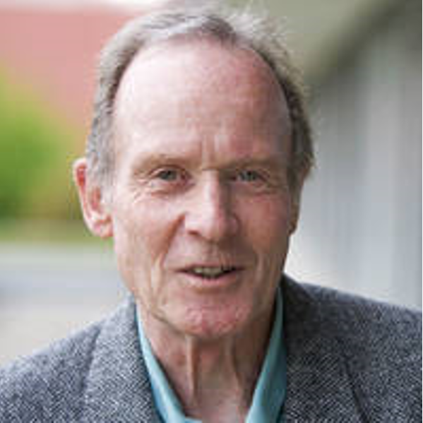
Microbial Rhodopsins are widely used in these days as optogenetic tools in neuro and cellbiology. We were able to show that rhodopsins from the unicellar alga Chlamydomonas reinhardtii with the 7 transmembrane helix motif act as light-gated ion channels, which we named channelrhodopsins (ChR1, ChR2). Together with the light-driven Cl-pump Halorhodopsin, ChR2 is used for the non-invasive manipulation of excitable cells and living animals by light with high temporal resolution and more important with extremely high spatial resolution. The basic functional mechanism and structural description of this unusual class of ion channels is given (electrophysiology, noise analysis, flash photolysis, and 2D crystallography and a high resolution structure of the WT and a mutant). Structure-based new tools and their application with a biomedical perspective in the cochlea and the restoration of vision are presented. |
|
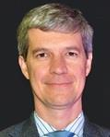
When coherent waves propagate through tissue that scatters the wave, it produces a speckle pattern. When the particles that scatter the coherent waves is moving, it will cause the speckle pattern to fluctuate in time. This is the basis of Laser Doppler blood flowmetry, diffuse correlation spectroscopy, laser speckle contrast imaging, and optical coherence tomography methods of measuring blood flow. The temporal fluctuations of the speckle intensity are generally described by the temporal auto-correlation function, which generally exhibits an exponential decay with temporal lag that goes as the mean square displacement of the scattering particles. One would expect that the mean square displacement of the red blood cells is dominated by the linear motion of the flowing red blood cells. But surprisingly experimental results by diffuse correlation spectroscopy 20 years ago indicated that the mean square displacement of the red blood cells was exhibiting a diffusive behavior. By combining measurements from diffuse cor- relation spectroscopy, optical coherence tomography and high-speed laser speckle contrast imaging, we unravel the mystery of this old unexplained behavior and establish that the shear induced diffusion of red blood cells is the dominant dynamics of measured by laser Doppler, diffuse correlation spectroscopy, and laser speckle contrast imaging, and is also a significant contributor to optical coherence tomography measurements of blood flow. If time permits, I will also describe how these speckle dynamics concepts can also be applied to new high-speed ultrasound measurements of blood flow dynamics. |
|
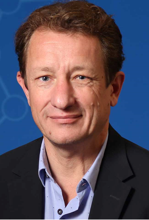
Patrick Degenaar It typically takes two decades to bring a new drug candidate or device concept to clinical practice. Optogenetics - a gene therapy technique to engender light sensitivity in the nervous system is 16 years old and now being actively explored in clinicaltrials for retinal prosthetics. A key question is whether and how it can be utilised in the brain for applications such as visual brain prosthetics and seizure suppression in epileptics. There are significant challenges in achieving this, from both technical and regulatory perspectives. In this talk I will present our technical progress with a particular focus on visual prosthesis, and our timeline to clinical. |
|
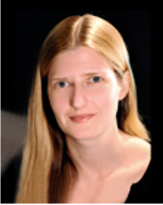
Elizabeth Hillman The past decade has seen dramatic improvements in genetically-encoded reporters of neural activity. However, capturing this activity at high speeds, over large volumes of the in intact brain and nervous system has remained a significant challenge. One technology that we have developed to address this problem is swept confocally aligned planar excitation (SCAPE) microscopy for high-speed 3D microscopy. SCAPE is a type of light sheet microscopy, but utilizes a novel scanning-descanning strategy to enable very high-speed volumetric cellular imaging with a versatile single, stationary objective at the sample. We are applying SCAPE to imaging awake, behaving organisms such as freely crawling Drosophila larvae, the whole brain of behaving adult Drosophila, zebrafish brain and the awake mouse cortex. We have also developed wide-field optical mapping (WFOM) for imaging both neural activity and hemodynamics over the entire dorsal cortical surface in awake, behaving mice. Both of these techniques are providing new high-speed, real time views of brain-wide activity in awake, behaving animals, providing fundamentally new views of spontaneous activity and behavior. I will present our latest progress on high-speed imaging technique development, and showcase our work applying these techniques to understand whole-brain activity in the context of awake behavior and resting state networks. |
|
All topics in neuroscience are informed by knowledge of brain cell type anatomy. The distribution and ratios of individual neuronal and glia cell types across the whole brain and the wiring of neuronal cell types into local and long-range circuits underlie the vast diversity of mammalian behaviors, from the simple startle response of defensive behaviors to the complex neuronal computations during cognitive and emotive processing. Here I will describe our work on systematic atlasing of cell-type distribution and morphology in C57BL/6 female and male mouse brain, including establishing novel microscopy methods for rapid imaging of mouse brains at high spatial resolution, computational methods for mapping cell-type distribution and tracing cellular morphologies, computational methods for registering the whole-brain datasets onto the Common Coordinate Framework (CCF) for postnatal day 56 (P56) mouse brain, statistical methods for rigorous analyses of the generated data, and neuroinformatics methods and an online web portal for integrating and disseminating the whole-brain cell type-based anatomical data. |
|
Dr. Sharan will present the application of lasers that are currently used in Neurosurgery such as the Visualase Laser System (Medtronics). He will also discuss current challenges and unmet needs in neurosurgical procedures that could be addressed by biophotonics. |
|
Neuromodulation has the potential to treat various diseases (i.e., heart failure, obesity). Using devices to modulate activity and treat diseases has great potential and may have several advantages compared to pharmaceutical approaches including cheaper costs to develop and less side effects. In the past, electric current has been primarily used for neuromodulation. Electric current has great promise, but there is also a need to develop other neuromodulation technologies that have different selectivity or other advantages. For example, the inability to modulate small-diameter axons was considered a critical factor of several clinical trial failures. Previously, we demonstrated unique selectivity (e.g., preferential inhibition of small-diameter fibers, high spatial selectivity) of peripheral structures (e.g., nerve, ganglia) using infrared neuromodulation (IRN). By using different protocols, we can either inhibit of stimulate activity. Here, we will present our recent developments which includes work on understanding the mechanisms and demonstrations of unique selectivity in a variety of tissues. |