
FEL Core Facilities
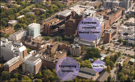
The core facilities of the Biophotonics Center is located at a renovated state-of-the-art facility, formerly known as the W.M. Keck Free Electron Laser (FEL) Center. This facility is located across from the Biomedical Engineering Department as well as other Science and Engineering buildings and within 300 yards of the Vanderbilt Medical Center. After the FEL program was ended the laboratory facilities underwent a $1 million renovation completed in August 2010 which resulted in over 5,000 square foot of state-of-the-art Biophotonics Center space. Set up in part as core labs and in part as thematic research labs, this new facility includes laboratories for clinical spectroscopy/diagnostics, Raman spectroscopy, optical imaging, Optical Coherence Tomography (OCT), neurophotonics, high power lasers as well as core support facilities that include bioluminescence imaging, spectrophotometry, image analysis/processing, microscopy, small animal surgery and fully equipped labs for cell / tissue culture, histology and an electronics / machine shop. Finally, the facilities include a state-of-the-art optics teaching lab to provide our undergraduate and graduate student with hands-on training in biomedical photonics. All laboratories are equipped with black-out curtains and each lab is partitioned into independent sections each with dedicated lighting and air handling systems.
The following are core facilities available at the FEL and do not include satellite facilities maintained by each individual faculty member:
Each faculty member has a faculty office in the BME department. Cubicle space for Postdoctoral fellows and Graduate Students is available in the Biophotonics Center and the BME Department on the 8th and 9th floor of the Science & Engineering building immediately adjacent to the FEL. Each person in the department has a personal computer linked to the VU network. Centralized data storage and backup facilities are provided and supported by a server in the BME department. Administrative support is available in the BME department. A large conference room (30+ people capacity) with state-of-the-art video conferencing and data sharing capabilities is available in the Biophotonics Center.
Vanderbilt University is an AAALAC accredited institution. Extensive animal care facilities are available to the investigators. This includes animal housing and care as well as a transgenic core facility maintained in the Vanderbilt-Ingram Cancer Center. The Animal Care Facility at Vanderbilt University Medical Center has four full time veterinarians (two of whom are ACLAM Diplomates) who are on call 24 hours a day for assistance with veterinary problems. The Biophotonics Center has in-house capabilities for surgeries on small animals including surgical tools, an operating microscope and two animal anesthesia systems (VetEquip).
The third floor of the Vanderbilt University Institute for Imaging Sciences (VUIIS) building houses various imaging systems operated by the Center for Small Animal Imaging (CSAI). Multimodal imaging capabilities include microPET, bioluminescence, microCT and several small animal MRI systems. Infrastructure and personnel for animal imaging support including surgery, anesthetics and animal handling are adjacent to the CSAI suite. In addition, wet laboratories for chemistry and cell culture laboratories are also located on this level. The CSAI is fully staffed with support personnel.
The Vanderbilt Medical Center is approximately 300 yards away from the Biophotonics Center. The clinical operating room is approximately 400 yards away and is easily accessible with a cart containing the systems. The Vanderbilt-Ingram Cancer Center and the Monroe-Carell Childrens’s Hospital are a little further at 500 yards. A state of the art clinical facility housing several clinics such as the Vanderbilt Women Center, Dermatology clinic etc.is located 10 miles away with dedicated space for research studies.
Every person in the Biophotonics Center has his or her own computer. In addition, there are PC based data acquisition systems available. Dedicated computers for instrument control (through LabVIEW), data collection and data analysis. There are several computers (PCs and a Macintosh) available in the Simulation and Computation modeling lab with various necessary software such as Matlab and Zeemax. Simulation for optical and thermal modeling is also accessible through these systems. Access is also available to high computing clusters (ACCRE), university servers and UNIX machines.
A large, competent electronics and machine shop is available for use with prior approval. This shop is equipped with machining tools such as micro drill press. Electronic and optical components are also available here.
The following equipment are available at the FEL:
- Microscopy: Fluorescence microscope (Zeiss, Axiovert 135) for UV and Visible wavelengths; Zeiss Stereo fluorescence microscope (SteREO Lumar); Zeiss stereo microscope (Discovery V08); Zeiss operating microscope, Moeller-Wedel operating microscope, Renishaw Ramascope (confocal Raman microscope)
- Lasers and other light sources: Q-switched Nd:YAG laser (Cobra); Nitrogen-dye laser (LaserPhotonics, LN1000/LN107), clinical CO2 laser; (3x) Schwartz Electro Optics high power pulse infrared laser (Holmium:YAG, Erbium:YAG and Nd:YAG); 1.85 μm diode laser (Aculight); 1.875 μm, diode laser (Aculight Capella); 1.94 μm diode laser (Aculight), He-Ne lasers; Xe-Hg arc lamp sources; deuterium lamp sources, NIST calibrated tungsten lamp
- Small animal imaging: Calcium (and intrinsic fluorescence) imaging system; hemodynamic imaging system; Bioluminescent Imaging system (Xenogen IVIS 100);Access to MRI and PET small snimal imaging systems through the Imaging Institute.
- Clinical diagnostic systems: Clinical fluorescence/diffuse reflectance spectroscopy systems; clinical hyperspectral fluorescence imaging system (home-built, LCTF-based); (4) clinical Raman spectroscopy systems (probe-based); clinical confocal imaging Raman spectroscopy system (home-built); clinical spatially offset Raman system, clinical combined Raman-OCT system,
- Other spectroscopy/imaging system: Benchtop fluorescence/diffuse reflectance system;,spectral imaging system (Applied Spectral Imaging); Raman microscope (Renishaw); benchtop combined Raman-OCT system; Optical Coherence Tomography (OCT) system (home-built); OCT system and hand-held imaging probe (Bioptigen), Swept Source Laser (Volcano Corp)
- Other relevant equipment: UV-Vis-NIR Spectrophotometer (Perkin-Elmer, Lambda900); Double integrating sphere setup for optical property measurements; Ocean Optics spectrometers; variety of optics and optics hardware; fiberoptics and related hardware; HeNe lasers; In house capability to build fiber probes; Raytracing software (ZeMax); computer-controlled translation stages; detectors (Molectron/Ophir/Newport); Digital storage oscilloscopes (4 Ch / 2 GHz) (3), function generators; amplifiers; video and imaging equipment, ultra-low temperature freezer; cell and tissue culture facilities; facilities for surgical procedures on small rodents (mice, rats); in house histology capabilities (incl. cryotome); various data acquisition systems; 13 vibration isolated optical tables; miscellaneous optics hardware. Tektronix TDS420 Digitizing Oscilloscope; NBS CO2 incubator; Revco Ultralow temp freezer; coldroom; Olympus BH2/CK2 microscopes, Nikon microscope, Leitz Laborlux microscope, and other general laboratory equipment.
-
Electrophysiology: stimulation/recording system consisting of: data acquisition system (Axon CNS, Digidata 1440A); Grass stimulator (S88x Astromed Inc.); Differential AC amplifier (model 1700, A-M Systems Inc.); Neuroprobe amplifier (model 1600, A-M Systems Inc.); vibration isolated Faraday cage (TMC); operating microscope (Zeiss); animal anesthesia system (VetEquip); salt water aquarium (for Aplysia); Clinical electrophysiology system (Endeavor, Nicolet Biomedical); DBS implant system (Radionics Stereoplan system; 4 Stealth Station treatment guidance Platforms; CRW stereotaxic system/adaptors; ACUSTAR system & frameless DBS implant system); human microelectrode recording system (4-ch Leadpoint x2, 10-ch Guideline 4000), accelerometer module model 2210; Nicolet Viking IV; WPI Isostem A320 microstimulators; Koph 650 microdrive; Grass S88 stimulator; WPI DAM 80 AC Differential Amplifier; Grass ICP511 AC Amplifier; Activa DBS quatropolar DBS lead #3387/#3389; Activa external stimulator 3625; rat stereotatic frame; Vibratome Series 1000 sectioning system; Sutter Puller P-2000; Narishige hydraulic micropositioner; interfaced chamber; Axopatch-1 D; air table; pCLAMP 8/Digidata 1320A data acquisition & analysis system.
The Biophotonics Center also has access to Vanderbilt University and Medical Center (VUMC) resources including the Center for Small Animal Imaging at the Institute of Imaging Science (VUIIS), which maintains several MRI scanners, CT-scanners, intravital fluorescence systems (IVIS), and more.