Yu, Lining; Yin, Mengmeng; Deng, Ruining; Liu, Quan; Yao, Tianyuan; Cui, Can; Guo, Junlin; Wang, Yu; Wang, Yaohong; Zhao, Shilin; Yang, Haichun; & Huo, Yuankai. (2025). Glo-In-One-v2: Holistic identification of glomerular cells, tissues, and lesions in human and mouse histopathology. Journal of Medical Imaging, 12(6), 61406. https://doi.org/10.1117/1.JMI.12.6.061406
Segmenting structures and lesions inside kidney glomeruli usually requires expert nephropathologists to carefully examine tissue morphology, a process that is time-consuming and can vary between observers. Building on their earlier Glo In One toolkit for detecting and segmenting glomeruli, the authors developed Glo In One version 2, which adds more detailed segmentation capabilities. They created a large annotated dataset containing 14 labels that cover tissue regions, cell types, and glomerular lesions across 23,529 glomeruli from both human and mouse kidney histopathology images, making it one of the largest datasets of its kind. Using this dataset, they trained a single deep learning model with a dynamic head architecture to segment all 14 classes from partially labeled whole slide images. The model was trained on 368 annotated kidney slides and learned to identify five intraglomerular tissue types and nine lesion types. The model achieved solid performance, with an average Dice similarity coefficient of 76.5 percent for glomerulus segmentation. In addition, using transfer learning, where knowledge learned from mouse data is applied to human data, improved lesion segmentation accuracy by more than 3 percent across lesion types. Overall, this work introduces a publicly available convolutional neural network that enables detailed, multiclass segmentation of glomerular tissue and lesions, helping reduce manual workload and variability in kidney pathology analysis.
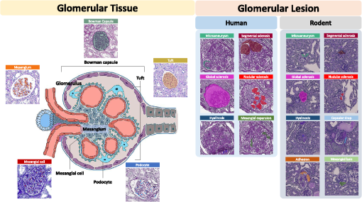
Fig. 1
This figure presents fine-grained classes of intraglomerular tissue, including Bowman’s capsule (Cap), tuft (Tuft), mesangium (Mes), mesangial cells (Mec), and podocytes (Pod). It also highlights the glomerular lesions observed in rodents and humans: AH, adhesion; CD, capsular drop; GS, global sclerosis; HS, hyalinosis; ML, mesangial lysis; MA, microaneurysm; NS, nodular sclerosis; ME, mesangial expansion; SS segmental sclerosis.