Schilling, Kurt G.; Teh, Irvin; Cohen-Adad, Julien; Dortch, Richard D.; Ibrahim, Ibrahim; Wang, Nian; Damon, Bruce M.; Cochran, Rory L.; & Leemans, Alexander. (2026). Diffusion tractography outside the brain: The road less travelled. Brain Structure and Function, 231(1), 7. https://doi.org/10.1007/s00429-025-03062-9
Diffusion tractography is an MRI technique that shows how fibers are organized inside tissues by tracking the movement of water molecules. It has been used mostly to study the white matter pathways in the brain, but researchers are now applying it to other parts of the body. This review looks at how tractography is being used in organs and tissues outside the brain, including the heart, spinal cord, peripheral nerves, kidneys, muscles, and prostate. Each of these areas requires adjustments in how the images are collected and analyzed because they move, have different tissue properties, or contain more complex structures than brain tissue. Although there are still challenges, such as dealing with motion during scanning and interpreting fiber patterns in tissues with less organized structure, tractography outside the brain offers a valuable, noninvasive way to study how tissues are organized at a microscopic level. These advances open new possibilities for both biomedical research and clinical care.
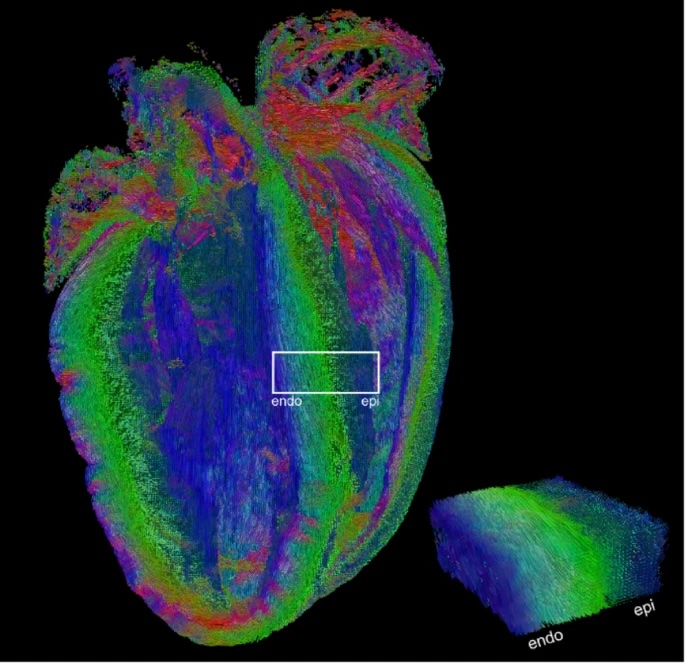
Fig. 1
Tractography of ex vivo mouse heart in 4-chamber view acquired at 35 μm isotropic resolution illustrates the transmural transition in orientation of cardiomyocytes in the left ventricular septal wall from subendocardium to subepicardium (highlighted white); underlying data from (Teh et al. 2024). Tracks are coloured according to directions: along the heart long-axis (blue), anterior-posterior (green) and septal-lateral directions (red)