Niu, Yanbin; Camacho, Maria Catalina; Schilling, Kurt G.; Humphreys, Kathryn Leigh. (2025). In vivo mapping of infant brain microstructure with neurite orientation dispersion and density imaging. Brain Structure and Function, 230(8), 147. https://doi.org/10.1007/s00429-025-03007-2
Diffusion magnetic resonance imaging (dMRI) is a non-invasive brain imaging technique that tracks the movement of water molecules in tissue over time. Because water movement is influenced by tiny cellular structures like membranes, axons, and myelin, dMRI provides a unique way to study the brain’s microstructure. One advanced dMRI method, called neurite orientation dispersion and density imaging (NODDI), models how brain cells and their connections are organized, giving detailed insights into tissue structure.
The early postnatal period is a time of rapid brain growth, including axonal growth, dendritic branching, and synapse formation. These processes change the brain’s microstructure in ways that NODDI can detect, making it a promising tool for studying early brain development. This review highlights recent studies using NODDI in infancy, showing how it can map typical developmental patterns, examine changes in preterm infants, and link microstructural properties to environmental factors and early behaviors.
While research is still limited—often with small sample sizes, narrow age ranges, and few longitudinal studies—initial findings suggest that NODDI can complement traditional diffusion measures and offer new insights into early neural development and brain plasticity. Continued use and refinement of NODDI in infants may help identify sensitive periods in brain development and improve understanding of emerging neurobehavioral traits.
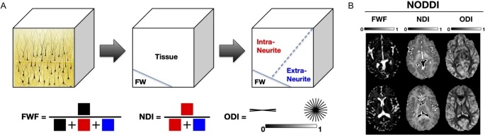
Fig 1
NODDI model components and representative maps of NODDI parameters. A The brain microstructure is modeled as three compartments: free water (FW), intra-neurite, and extra-neurite spaces. The free water fraction (FWF), neurite density index (NDI), and orientation dispersion index (ODI) are derived from these compartments. B Representative axial slices from NODDI-derived maps for FWF, NDI, and ODI. Note: Figure adapted from Kraguljac et al. (2023), licensed under Creative Commons Attribution-NoDerivatives 4.0 International (CC BY-ND 4.0), available at http://mig.cs.ucl.ac.uk/index.php?n=Tutorial.NODDImatlab