Witt, Atlee A., Combes, Anna J.E., Sweeney, Grace, Prock, Logan E., Houston, Delaney C., Stubblefield, Seth K., McKnight, Colin David, O’Grady, Kristin P., Smith, Seth A., & Schilling, Kurt G. (2025). Leveling up: along-level diffusion tensor imaging in the spinal cord of multiple sclerosis patients. Frontiers in Neuroimaging, 4, 1599966. https://doi.org/10.3389/fnimg.2025.1599966
Multiple sclerosis (MS) is a chronic disease of the nervous system that causes inflammation, damage to the protective covering of nerve fibers (demyelination), and degeneration of axons. These changes can be studied using diffusion tensor imaging (DTI), which measures microstructural damage in the brain and spinal cord. In the brain, researchers often use white matter (WM) tractography to examine changes along specific pathways. In the spinal cord (SC), however, anatomy is naturally divided into cervical levels, which provides a different way to study regional changes.
In this study, we used an along-level approach to measure both microstructural features (such as fractional anisotropy, a DTI measure of tissue integrity) and macrostructural features (such as cross-sectional area) of the SC in people with relapsing-remitting MS (pwRRMS) compared to healthy controls (HCs).
The results showed that analyzing the SC level by level was more sensitive to detecting group differences than averaging across the whole cord. Segmenting the cord into WM tracts and gray matter (GM) subregions revealed specific, localized changes along the cord and within its cross-sections. Importantly, GM atrophy was linked with greater clinical disability, whereas microstructural changes did not show significant associations with disability measures.
These findings highlight the value of level-specific analysis for identifying localized spinal cord pathology and suggest a more refined framework for studying SC changes in MS.
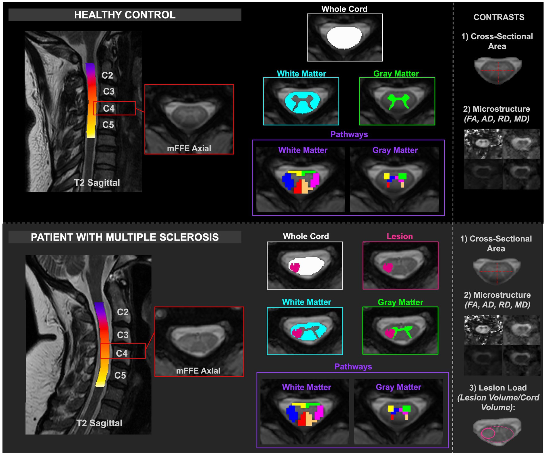
Figure 1. Depiction of healthy and MS cord processing, including delineation of the masks relative to healthy or lesioned tissue. The contrasts are included in the right column. CSA and diffusion-derived indices were calculated for HCs, and CSA, diffusion-derived indices, and lesion load were calculated for pwRRMS.