Fan, Jiawen, Juttukonda, Meher R., Goodale, Sarah E., Wang, Shiyu, Orban, Csaba, Varadarajan, Divya, Polimeni, Jonathan R., Chang, Catie E., Salat, David H., & Chen, Jingyuan E. (2025). Functional MRI signatures of autonomic physiology in aging. Communications Biology, 8(1), 1287. https://doi.org/10.1038/s42003-025-08703-7
In brain imaging research, small changes in functional MRI (fMRI) signals caused by breathing and heart rate have often been dismissed as “noise.” However, these fluctuations actually contain important information about how blood vessels in the brain and the body’s autonomic functions (like heart and breathing control) work.
In this study, we used these physiological signals to examine how brain function changes with age, using data from the large Lifespan Human Connectome Project Aging study. We found that as people get older, their fMRI signals show slower responses linked to breathing, faster responses linked to the heartbeat, and stronger connections between brain and heart signals. Importantly, these changes become especially noticeable after the age of 60, suggesting that declining vascular health and changes in autonomic function play a key role in aging.
We also tested whether these fMRI patterns might be influenced by age-related changes in brain structure, blood flow, or alertness during scans. Overall, our findings highlight major age effects in fMRI signals tied to heart and breathing activity. This work shows that resting-state fMRI can be used not only to study brain connectivity but also to reveal new markers of brain physiology that could help track vascular and autonomic changes with aging.
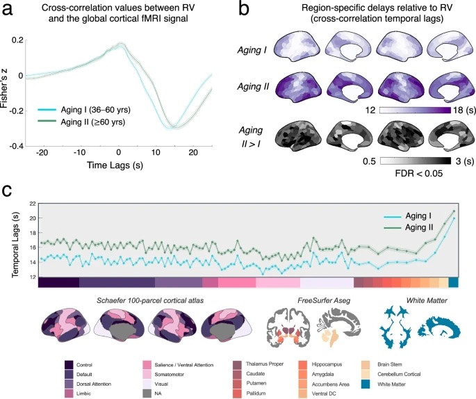
Fig. 1: Age effects on the spatiotemporal patterns of RV-coupled fMRI dynamics.
a The cross correlation between the global cortical fMRI signal and RV (positive lag values suggest that fMRI signals lag RV) for different age groups, with shade denoting the standard errors across subjects. b Intra-cortical distributions of region-specific fMRI signal lag relative to RV (based on the Schaefer 300-parcel atlas). Regions exhibiting statistically significant between-group temporal lag differences were displayed at the bottom (“Aging II > I”, FDR < 0.05). c Tissue-type specific temporal lags relative to RV for each age group. Error bars indicate the standard errors of RV-fMRI temporal lags across subjects, and the shaded gray area highlights ROIs that exhibited statistically significant between-group differences (FDR < 0.05). ROI labels are shown at the bottom.