Ding, Zhaohua, Xu, Lyuan, Gao, Yurui, Zhao, Yu, Tan, Yicheng, Anderson, Adam W., Li, Muwei, & Gore, John C. (2025). Cortical modulation of resting state BOLD signals in white matter. Scientific Reports, 15(1), 30056. https://doi.org/10.1038/s41598-025-14352-x
Magnetic resonance images of healthy brains were analyzed to better understand how resting-state BOLD signals in white matter are related to neural activity in the cortex (the outer layer of the brain). We measured how much spontaneous activity in the cortex—seen as low-frequency fluctuations in BOLD signals from gray matter—affects the resting-state BOLD signals in white matter. We found that the similarity between BOLD signals from cortical regions and white matter areas was directly linked to the strength of the cortical BOLD signal.
From these measurements, we observed that cortical networks involved in more basic functions tend to contribute more to the fluctuations in white matter than those involved in higher-level functions. We also discovered that each cortical network has its own unique spatial pattern of influence on white matter BOLD signals, and the strength of these effects is closely related to how much myelin (the protective coating around nerve fibers) the cortical network has.
Overall, our findings show that resting-state BOLD signals in white matter reflect the spontaneous activity of specific cortical networks and are shaped by the structure and myelination of the cortex.
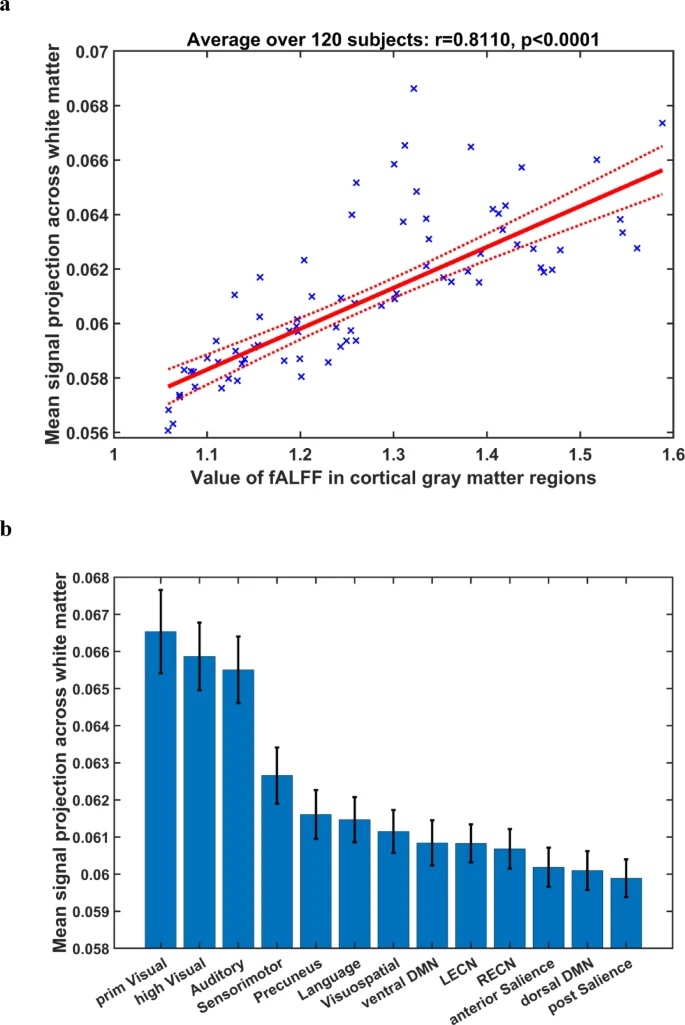
Fig 1
(a) Relationship between subject-averaged fALFF of cortical BOLD signals and their subject-averaged mean white matter projection. Each data point represents subject-averaged measures for an ROI in the cortex. (b) Mean white matter projection of BOLD signals in the cortical functional networks analyzed. The vertical line at the top of each bar represents standard error across the 120 subjects studied. Abbreviations: prim = primary, DMN = default mode network. LECN = left executive control network. RECN = right executive control network.