Song, Richard, Min, Jungwon, Wang, Shiyu, Goodale, Sarah E., Rogge-Obando, Kimberly K., Yang, Ruoqi, Yoo, Hyunjoo, Nashiro, Kaoru, Chen, Jingyuan E., & Mather, Mara M. [2025]. “The physiological component of the BOLD signal: Impact of age and heart rate variability biofeedback training.” Imaging Neuroscience, 3, IMAG.a.99. The physiological component of the BOLD signal: Impact of age and heart rate variability biofeedback training
Aging is linked to declines in the autonomic nervous system [which controls things like heart rate and breathing], reduced coordination between brain activity and blood flow, and weaker blood vessel responses. These changes may play a role in memory loss and neurodegenerative diseases. To better understand this, we studied how aging affects the way the brain integrates signals from the heart, lungs, and blood flow.
Using two independent brain imaging [resting-state fMRI] datasets with heart and breathing measurements from younger and older adults, we found that older adults showed reduced connections between heart rate, breathing patterns, carbon dioxide levels, and the brain’s oxygenation signal [BOLD signal]. These reductions were most noticeable in brain regions that help regulate automatic body functions, such as the orbitofrontal cortex, anterior cingulate cortex, insula, basal ganglia, and white matter. Younger adults showed stronger heart rate–brain signal coupling in white matter and faster brain responses to breathing and carbon dioxide changes in gray matter.
We also tested whether heart rate variability biofeedback [HRV-BF]—a non-invasive breathing-based training that improves natural heart rate rhythms—could affect these brain-body connections. In older adults, HRV-BF shifted heart rate–brain coupling patterns to look more like those of younger adults.
These results suggest that HRV-BF may help counteract age-related declines in brain and blood vessel function. Overall, this study shows how closely linked body rhythms are to brain health and highlights a potential strategy to support brain function and preserve cognitive health as we age.
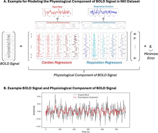
Fig 1 – Schematic for the model to determine the physiological component of the BOLD signal. [A] After detrending and normalizing heart rate and respiratory variation, the signals are convolved with CRF and RRF basis functions. For every voxel, a general linear model is used to find beta weights for each of the cardiac and respiration regressors to minimize the error from the original BOLD signal. [B] Example of an original BOLD signal [normalized to percent signal change] and the corresponding physiological component.