Panzer, Stephanie, Wörtler, Klaus, Paladin, Alice, Zesch, Stephanie, Rosendahl, Wilfried, van Schaik, Katherine D., Sutherland, M. Linda, Sutherland, James D., Hergan, Klaus, Thompson, Randall C., & Zink, Albert R. (2025). Systematic assessment of bone and soft tissue tumors on whole-body CTs of 45 mummies from ancient Egypt. *Scientific Reports, 15*(1), 21482. https://doi.org/10.1038/s41598-025-07029-y
There is growing interest in how long cancer has existed and why malignant tumors, especially in soft tissues, seem rare in ancient human remains. To explore this, researchers carefully examined 45 whole-body CT scans of ancient Egyptian mummies to look for bone and soft tissue tumors. They found evidence of malignant bone disease (likely cancer spread to the bones) in 1 out of 45 cases (2%). In addition, 5 out of 45 mummies (11%) showed soft tissue masses that were likely cancerous. These soft tissue tumors had clear edges, different internal patterns, and were denser than the nearby preserved soft tissues. In two cases where soft tissue tumors were inside the abdomen, the original organs were not preserved. In summary, malignant tumors, including those in soft tissues, can be detected using CT scans of ancient Egyptian mummies. This discovery about how these tumors appear and how often they occur provides new information and a fresh way to study cancer in ancient populations.
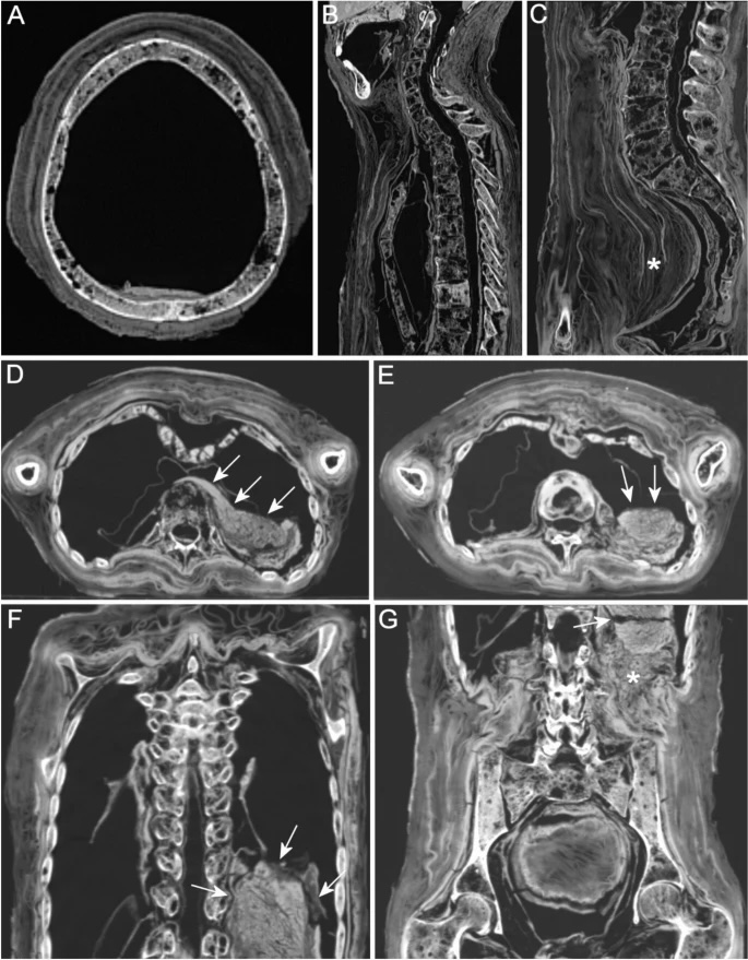
Fig 1
Case 16, probable skeletal metastases and large intra-abdominal soft tissue mass. (A) Axial multiplanar (MPR) reconstruction of the skull illustrating multiple predominantly small osteolytic lesions of the cranial vault involving the outer and inner table as well as the diploe. (B) Sagittal MPR of the cervical and thoracic spine demonstrating multiple osteolytic lesions in the vertebral bodies, spinous processes and the sternum. The second thoracic vertebra reveals a healed burst fracture with collapse, the first and third thoracic vertebrae show infraction of the adjacent endplates. (C) Sagittal MPR of the lumbar spine and the sacrum showing multiple osteolytic lesions. Note the cachectic body and the textiles that overly the lumbar spine and protrude into the relatively empty pelvic cavity (asterisk). (D) Axial MPR of the upper abdomen illustrates a large soft tissue mass that expands from the midline towards the left dorsolateral part of the abdomen (arrows). In the midline, the mass is relatively homogeneous, in the lateral part, it shows different components/layers with stratified, loosened structure. The mass appears to remodel the pancreas body and tail. (E) Axial MPR of the middle abdomen demonstrating the lower part of the mass with different components/layers (arrows). (F) Coronal MPR of the upper abdomen illustrating the soft tissues mass with irregular contour of the upper margin (arrows). Note preservation of shrunken lungs bilaterally. Unfortunately, the CT scan was sectioned at the level of the soft tissue mass. (G) Coronal MPR of the middle and lower abdomen showing the mass with a horizontal postmortem split due to desiccation (arrow). There is increased adjacent soft tissue around the lower margin (asterisk).