Cho, Chloe, Chamberland, Maxime, Rheault, François, Moyer, Daniel, Landman, Bennett A., & Schilling, Kurt G. (2025). Microstructural characterization of short association fibers related to long-range white matter tracts in normative development. *Human Brain Mapping, 46*(8), e70255. https://doi.org/10.1002/hbm.70255
Short association fibers (SAFs) are small nerve fibers located in the outer layers of the brain’s white matter that help nearby regions of the brain communicate with each other. Although they play a key role in local brain connections, SAFs have not been widely studied until recently, due to the lack of imaging methods able to capture this part of the brain in detail. Understanding how SAFs develop, especially in comparison to longer white matter tracts that connect distant brain regions, is important for learning how the brain matures from childhood through young adulthood.
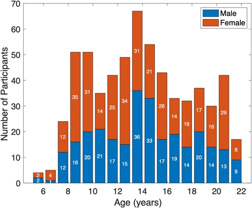
This study set out to do three things: map where SAFs are located in relation to long-range white matter tracts, describe how these fibers change during normal brain development, and explore how changes in SAFs relate to changes in the long-range connections. Researchers studied brain scans from 616 participants, ages 5.6 to 21.9 years old, using advanced imaging methods called diffusion tensor imaging (DTI) and neurite orientation dispersion and density imaging (NODDI). These techniques allow scientists to look at how brain tissue is structured and how water moves through it, which provides insights into how brain connections grow and change.
The results showed that both SAFs and long-range white matter tracts followed similar patterns of development. Some features, such as the way water diffuses in the brain (measured by MD, AD, and RD), decreased with age, while other features (like FA, ICVF, ISOVF, and ODI) increased, which is consistent with healthy brain development. However, SAFs and long-range tracts also showed important differences in their development, suggesting that the outer and deeper parts of the brain’s white matter mature in slightly different ways.
In addition to age-related findings, the study also found differences between males and females in several brain measures. These findings highlight that brain development can vary by sex. Overall, this study offers new insights into how the brain’s short- and long-range connections grow during childhood and adolescence, and provides a strong foundation for future research into unusual brain development or disease.
Figure 1
Age and sex distribution in the study cohort of 616 participants (279 M, 337 F), ranging from 5.6 to 21.9 years old with a mean age of 14.5 years.