Jones, Natalie; Schwartz, Trent M.; Bishay, Steven; Robb, W. Hudson; Sams, Amanda; Kogan, Josh; Nable, Monica; Nelson, Sydney; Zhao, Oliver S.; Hohman, Timothy; Huang, Steven; Martinez, Felipe; Nguyen, Ba; Shin, Clifford; Yang, Ming; Westervelt, Holly; Szymkowicz, Sarah M.; Omary, Lesley T.; Aqel, Bashar A.; Dickson, Rolland; Lizaola, Blanca; Mathur, Amit; Izzy, Manhal; Koran, Mary Ellen I. “FDG PET of the brain to screen for neurodegenerative disease in older liver transplant candidates.” European Journal of Nuclear Medicine and Molecular Imaging (2025). https://doi.org/10.1007/s00259-025-07382-0.
As more older adults become candidates for liver transplants, it’s increasingly important to distinguish between dementia caused by aging and hepatic encephalopathy (HE)—a condition related to liver dysfunction. These two conditions can appear very similar in how they affect thinking and memory. While neuropsychological testing (NPT) is commonly used to evaluate dementia, imaging techniques like FDG PET/CT scans, which measure brain activity, are being explored as faster alternatives. This preliminary study looks at how useful FDG PET/CT is for detecting irreversible dementia in older people being considered for a liver transplant.
Eighteen patients showing signs of possible dementia during their transplant evaluations received an FDG PET/CT brain scan. The study compared the PET/CT scan results to NPT results and also looked at how long each test took to produce a diagnosis. Brain scan results were also compared to healthy individuals of the same age to see if there were noticeable differences in brain activity.
In 40% of the patients, the PET/CT scan identified signs of irreversible dementia. The scan results moderately agreed with the NPT results, and while the scan was highly sensitive (able to catch all cases of dementia), it wasn’t as specific (sometimes showing false positives). Importantly, the PET/CT scan delivered results much faster—on average in about 12 days, compared to over 70 days for NPT. The scans also revealed significant reductions in brain activity in areas linked to thinking and movement, compared to healthy individuals.
These findings suggest that FDG PET/CT scans could be a useful early tool in identifying irreversible causes of dementia in older liver transplant patients. However, a positive scan should not be the sole reason to rule someone out of receiving a transplant; it should lead to further testing. Using PET/CT scans as an initial screening step could speed up the evaluation process and help ensure that patients who need further care get it in time.
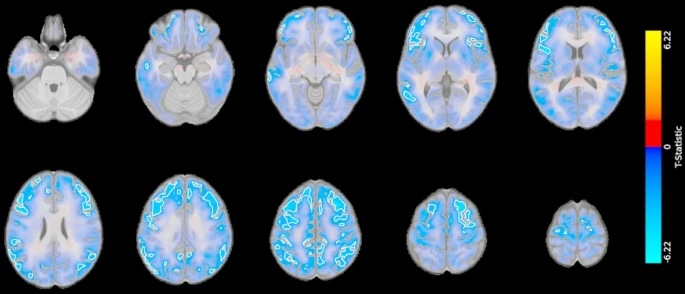
Fig. 1
Results from matched case-control comparison of FDG PET scans of the brain. Cases (patients referred for FDG PET for cognitive symptoms during transplant evaluation) had significantly decreased fluorodeoxyglucose (FDG) uptake in multiple large clusters (outlined in white) primarily in the frontal, temporal, and parietal lobes compared to healthy age- and sex-matched controls from the Alzheimer’s Disease Neuroimaging Initiative database (ADNI). These T-statistic maps show the regional statistical differences in glucose metabolism in the brain between 16 case-control pairs. Blue reflects voxels where cases had decreased FDG uptake vs. controls, while red reflects the opposite. Clusters that reach significance (FDR < 0.05 and voxel-extent of 10) are outlined in white. No significant clusters of increased SUVR were observed.