Mu, Chaoqi; Reed, Jamie L.; Wang, Feng; Yan, Xinqiang; Lu, Ming; Gore, John C.; Chen, Li Min. “Validation of qMT and CEST MRI as Biomarkers of Response to Treatment After Lumbar Spinal Cord Injury in Rats.” NMR in Biomedicine 38, no. 4 (2025): e70015. https://doi.org/10.1002/nbm.70015.
When the spinal cord is injured, the damage and healing processes are complex and involve many different biological changes. To better understand and track these changes, especially in studies that test treatments in animals before trying them in people, scientists are looking for reliable and noninvasive ways to measure what’s happening inside the body. This study focused on using a special type of MRI scan to find signals, or “biomarkers,” that reflect the severity of spinal cord injury, the loss and repair of protective nerve coverings (called myelin), and inflammation in the nervous system.
Researchers used two advanced MRI techniques—chemical exchange saturation transfer (CEST) and quantitative magnetization transfer (qMT)—to study rats that had a moderate spinal cord injury. Some of the rats were treated with a drug called riluzole, which may protect nerve cells, while others received a control treatment. Over the course of eight weeks, the scientists monitored changes in the rats’ spinal cords using these MRI methods and then compared the results to lab tests and how well the animals could move and feel.
They found that rats treated with riluzole had signs of better myelin repair in their spinal cords, and less inflammation, compared to untreated rats. These findings were backed up by tissue analysis after the animals were studied. The MRI measurements also matched up with how well the rats recovered, suggesting that the scans were picking up meaningful signs of healing.
Overall, this research shows that these advanced MRI techniques can be powerful tools for tracking spinal cord injury and recovery. They help researchers understand how treatments like riluzole are working, and could make it easier to apply what’s learned in animal studies to help people in the future.
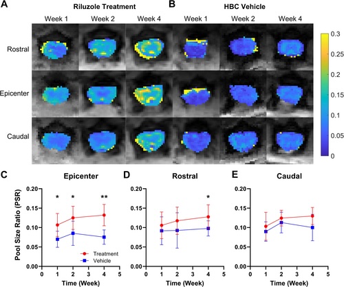
FIGURE 1
Comparison of qMT PSR maps and values along the spinal cord in riluzole-treated versus HBC vehicle-treated SCI rats. (A,B) Representative PSR maps acquired from injury epicenter, rostral, and caudal to the injury at Week 1 and Week 2 to Week 4 post-injury in rats that received riluzole (A) versus HBC vehicle (B) treatment. (C–E) Average white matter PSR values comparison between treatment and vehicle groups, at (C) injury epicenter, (D) rostral, and (E) caudal, from Week 1 to Week 4. *p < 0.05, **p < 0.005, non-parametric Wilcoxon rank-sum test.