Schilling, Kurt G.; Grussu, Francesco; Ianus, Andrada; Hansen, Brian; Howard, Amy F. D.; Barrett, Rachel L. C.; Aggarwal, Manisha; Michielse, Stijn; Nasrallah, Fatima; Syeda, Warda; Wang, Nian; Veraart, Jelle; Roebroeck, Alard; Bagdasarian, Andrew F.; Eichner, Cornelius; Sepehrband, Farshid; Zimmermann, Jan; Soustelle, Lucas; Bowman, Christien; Tendler, Benjamin C.; Hertanu, Andreea; Jeurissen, Ben; Verhoye, Marleen; Frydman, Lucio; van de Looij, Yohan; Hike, David; Dunn, Jeff F.; Miller, Karla; Landman, Bennett A.; Shemesh, Noam; Anderson, Adam; McKinnon, Emilie; Farquharson, Shawna; Dell’Acqua, Flavio; Pierpaoli, Carlo; Drobnjak, Ivana; Leemans, Alexander; Harkins, Kevin D.; Descoteaux, Maxime; Xu, Duan; Huang, Hao; Santin, Mathieu D.; Grant, Samuel C.; Obenaus, Andre; Kim, Gene S.; Wu, Dan; Le Bihan, Denis; Blackband, Stephen J.; Ciobanu, Luisa; Fieremans, Els; Bai, Ruiliang; Leergaard, Trygve B.; Zhang, Jiangyang; Dyrby, Tim B.; Johnson, G. Allan; Cohen-Adad, Julien; Budde, Matthew D.; Jelescu, Ileana O. “Considerations and recommendations from the ISMRM diffusion study group for preclinical diffusion MRI: Part 2—Ex vivo imaging: Added value and acquisition.” Magnetic Resonance in Medicine 93, no. 6 (2025): 2535-2560. https://doi.org/10.1002/mrm.30435.
Diffusion MRI (dMRI) is a powerful imaging method that lets scientists study the structure and connections of tissues, especially the brain. While it’s often used to scan living animals (called in vivo imaging), researchers are increasingly using it to scan tissues that have been removed from the body (ex vivo imaging). This type of imaging has some big advantages: it can produce clearer, more detailed images and allows for more advanced ways to study the tiny structures and connections in the brain. It also lets scientists directly compare MRI results with microscopic images of the same tissue, which helps confirm that their methods are accurate.
However, ex vivo imaging comes with its own challenges. Everything from how the tissue is prepared to how the images are taken and analyzed is more complex than scanning a live animal. These steps can strongly influence what kinds of questions researchers can answer.
This paper is the second in a three-part series that gives advice for using dMRI in research. In this part, the authors focus on best practices for scanning and analyzing tissue that’s been removed from the body. They cover the basics researchers need to know when planning experiments, explain different types of tissue samples and when to use each, and provide detailed advice on how to prepare the samples and run the MRI scans.
They also point out areas where there aren’t clear rules yet—and where more research is needed. Overall, the goal is to improve the quality and consistency of ex vivo dMRI so it can help push medical science forward.
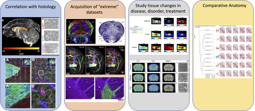
FIGURE 1
Four areas in which preclinical brain imaging adds value to the field of dMRI. It enables: (i) correlation with histology on the same subject/sample, (ii) the acquisition of richer datasets than on clinical systems thanks to more advanced hardware and longer scan times available, (iii) the study of tissue changes with disease and treatment in a more controlled setting, and (iv) comparative anatomy between species. Figures reused and adapted from (left to right): (i),3–5 (ii),6–8 (iii),9, 10 (iv).11