Jelescu, Ileana O.; Grussu, Francesco; Ianus, Andrada; Hansen, Brian; Barrett, Rachel L. C.; Aggarwal, Manisha; Michielse, Stijn; Nasrallah, Fatima; Syeda, Warda; Wang, Nian; Veraart, Jelle; Roebroeck, Alard; Bagdasarian, Andrew F.; Eichner, Cornelius; Sepehrband, Farshid; Zimmermann, Jan; Soustelle, Lucas; Bowman, Christien; Tendler, Benjamin C.; Hertanu, Andreea; Jeurissen, Ben; Verhoye, Marleen; Frydman, Lucio; van de Looij, Yohan; Hike, David; Dunn, Jeff F.; Miller, Karla; Landman, Bennett A.; Shemesh, Noam; Anderson, Adam; McKinnon, Emilie; Farquharson, Shawna; Dell’Acqua, Flavio; Pierpaoli, Carlo; Drobnjak, Ivana; Leemans, Alexander; Harkins, Kevin D.; Descoteaux, Maxime; Xu, Duan; Huang, Hao; Santin, Mathieu D.; Grant, Samuel C.; Obenaus, Andre; Kim, Gene S.; Wu, Dan; Le Bihan, Denis; Blackband, Stephen J.; Ciobanu, Luisa; Fieremans, Els; Bai, Ruiliang; Leergaard, Trygve B.; Zhang, Jiangyang; Dyrby, Tim B.; Johnson, G. Allan; Cohen-Adad, Julien; Budde, Matthew D.; Schilling, Kurt G. “Considerations and recommendations from the ISMRM diffusion study group for preclinical diffusion MRI: Part 1: In vivo small-animal imaging.” Magnetic Resonance in Medicine 93, no. 6 (2025): 2507-2534. https://doi.org/10.1002/mrm.30429.
Diffusion MRI (dMRI) is a special type of brain scan that researchers use to study the structure and connections inside the body. In small animals like mice and rats, it’s often used to test new imaging methods, understand how tissues behave, and compare anatomy across different species. However, doing these scans on live animals (in vivo) is complicated. From preparing the animal and choosing the right equipment, to taking the images and making sense of the data—each step involves choices that can affect what scientists are able to learn.
This paper shares expert recommendations on how to best perform in vivo dMRI in small animals. It explains the important things researchers need to consider when planning these experiments, such as the pros and cons of using different animal species or disease models. It also offers practical advice on how to prepare the animals, what types of scanners and settings to use, and how to process and analyze the images afterward.
The authors also created an online guide that lists free dMRI datasets and software tools to help promote careful and repeatable research. While the focus is mostly on the brain and nervous system, the paper also gives tips for using dMRI on other organs when possible.
Overall, the goal is to help scientists do high-quality, reliable dMRI research in animals, so we can better understand health and disease and move medical science forward.
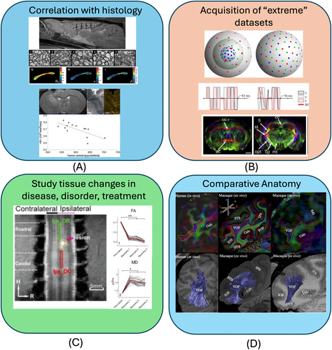
FIGURE 1
Four areas in which preclinical brain imaging adds value to the field of dMRI. It enables: (A) correlation with histology on the same subject/sample; (B) the acquisition of richer datasets than on clinical systems thanks to more advanced hardware and longer scan times available; (C) the study of tissue changes with disease and treatment in a more controlled setting; and (D) comparative anatomy between species. Figures reused and adapted from (A),10, 11 (B),12–15(C),16 (D).17