Hillyer Lab News

Article on the functional integration of the circulatory, immune and respiratory systems of larvae is published in BMC Biology
Monday, September 19, 2016
Insects have evolved powerful innate immune responses to neutralize infectious agents. Upon entering the hemocoel, cellular and humoral immune factors kill pathogens by phagocytosis, melanization, lysis, and other mechanisms. The primary immune cells that drive these processes are the hemocytes, which are found both circulating with the hemolymph and attached to tissues (sessile).
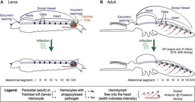
Immune responses in the hemocoel occur in a dynamic space, where hemocytes, humoral immune factors, and pathogens are propelled throughout the body by the action of an open circulatory system. This circulation is mediated by the contractile action of a muscular dorsal vessel that is anatomically divided into an abdominal heart and a thoracic aorta. Our earlier research has shown the functional integration of the circulatory and immune systems of adult mosquitoes, where cellular immune responses are rapid and inducible on the surface of the heart. Specifically, in adult mosquitoes, a population of sessile hemocytes, called periostial hemocytes, is always present on the surface of the heart in the regions that flank the heart valves, or ostia. At this location, periostial hemocytes phagocytose pathogens as they are swept with the hemolymph toward the heart. This capture of circulating pathogens induces the migration of additional hemocytes to the periostial region of each abdominal segment, where they aggregate with the initial population of first responders and amplify the phagocytosis response.
However, hemolymph circulation in adult and larval mosquitoes is drastically different, as, for example, the abdominal ostia of larvae are non-functional, and all hemolymph enters the heart via incurrent openings located at the posterior of the larva. In the present study we assessed whether the circulatory and immune systems of mosquito larvae interact during the course of an infection, and describe a pattern that is remarkably different from what is seen in adults. Specifically, we show that:
-
1.Unlike in adults, infection of a mosquito larva does not induce the aggregation of hemocytes and pathogens in the periostial regions of the heart.
-
2.Instead, infection induces the rapid aggregation and killing of pathogens on the surface of the tracheal tufts, which are respiratory structures that are unique to larvae and associate with the posterior terminus of the heart in the eighth abdominal segment.
-
3.In a process that is functionally analogous to what is seen in the periostial regions of adults, the tracheal tuft hemocytes of larvae phagocytose pathogens as they near entry into the dorsal vessel, and thus sequester and kill pathogens in the area of the body with the highest hemolymph flow.
Taken together, these findings show that the functional integration of the circulatory and immune systems of mosquitoes displays drastic stage-specific differences that reflect the broad changes in body plan that occur during metamorphosis.
Article citation:
League, G.P., and J.F. Hillyer. 2016. Integration of the circulatory, immune, and respiratory systems in mosquito larvae: immunity in the hemocyte-rich tracheal tufts. BMC Biology. 14:78. (PMID 27643786) (See it for FREE in BMC Biol)
Graphical abstract:
Article abstract:
Background: As both larvae and adults, mosquitoes encounter a barrage of immune insults, ranging from microbe-rich communities in larval habitats to ingested blood-borne pathogens in adult blood meals. Given that mosquito adults have evolved an efficient means of eliminating infections in their hemocoel (body cavity) via the coordinated action of their immune and circulatory systems, the goal of the present study was to determine whether such functional integration is also present in larvae. Results: By fluorescently labeling hemocytes (immune cells), pericardial cells, and the heart, we discovered that fourth instar larvae, unlike adults, contain segmental hemocytes but lack the periostial hemocytes that surround the ostia (heart valves) in abdominal segments 2–7. Instead, larvae contain an abundance of sessile hemocytes at the tracheal tufts, which are respiratory structures that are unique to larvae, are located in the posterior-most abdominal segment, and surround what in larvae are the sole incurrent openings for hemolymph entry into the heart. Injection of fluorescent immune elicitors and bacteria into the larval hemocoel then showed that tracheal tuft hemocytes mount rapid and robust immune responses against foreign insults. Indeed, green fluorescent protein-labeled Escherichia coli flowing with the hemolymph rapidly aggregate exclusively at the tracheal tufts, where they are killed within 24 h post-infection via both phagocytosis and melanization. Conclusion: Together, these findings show that the functional integration of the circulatory, respiratory, and immune systems of mosquitoes varies drastically across life stages.
According to the journal website, “BMC Biology is the flagship biology journal of the BMC series, pioneer of open access publishing. Our scope includes research and methodology articles, review, Q&A and comment, across all of biology and with a publication policy that combines selection for special interest and importance with a commitment to serving authors well.”