Hillyer Lab News

Article on periostial hemocytes and hemolymph flow is published in Developmental and Comparative Immunology
Monday, February 1, 2016
Mosquitoes have evolved robust innate immune responses that destroy pathogens. These immune responses are primarily driven by hemocytes, which are dedicated immune cells that can be found both circulating with the hemolymph as well as attached to tissues.
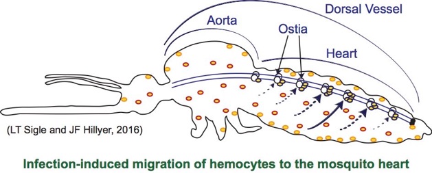
Because hemocytes, humoral immune factors and pathogens are present in the hemocoel, their biology is impacted by the circulation of hemolymph. Hemolymph in mosquitoes is propelled via the contractile action of a muscular tube called the dorsal vessel. This dorsal vessel extends the length of the body and is divided into two sections: the aorta in the thorax and the heart in the abdomen. Wave-like contractions of heart muscle propel the hemolymph, and these contractions periodically reverse the direction. When the contractions propagate toward the head (anterograde), hemolymph enters the dorsal vessel through 6 pairs of ostia (valves) that are sequentially located in the anterior portion of abdominal segments 2 through 7, and exits the vessel in the head. When the contractions propagate toward the posterior of the abdomen (retrograde), hemolymph enters the dorsal vessel through a single pair of ostia located at the thoraco-abdominal junction and exits the vessel in the 8th abdominal segment.
Recently we described a novel mosquito cellular immune response that occurs on the surface of the heart. Specifically, in the absence of infection, mosquitoes possess sessile hemocytes that are attached to the heart at the location of the ostia. These periostial hemocytes rapidly phagocytose pathogens that are circulating with the hemolymph, which is advantageous because these immune cells are in the areas of highest hemolymph flow, thus increasing the probability that they will encounter circulating pathogens.
In this study we scrutinized the immune response mounted by the periostial hemocytes of the malaria mosquito, Anopheles gambiae, against multiple bacterial pathogens. We found that:
-
1.Infection induces the aggregation of hemocytes in the periostial regions of the heart.
-
2.Periostial hemocyte aggregation is a basal immune response that is activated by diverse stimuli.
-
3.Periostial hemocyte aggregation is dynamic and preferentially occurs in the mid-abdominal segments.
-
4.Periostial hemocyte aggregation correlates with spatial patterns of immune activity and hemolymph flow.
Article citation:
Sigle, L.T. and J.F. Hillyer. 2016. Mosquito hemocytes preferentially aggregate and phagocytose pathogens in the periostial regions of the heart that experience the most hemolymph flow. Developmental and Comparative Immunology. 55:90-101. (PMID 26526332) (See it in DCI) (Email me for a pdf copy)
Graphical abstract:
Article abstract:
When a mosquito acquires an infection in the hemocoel, dedicated immune cells called hemocytes aggregate around the valves of the heart. These sessile hemocytes are called periostial hemocytes. In the present study we scrutinized the immune response mounted by the periostial hemocytes of the malaria mosquito, Anopheles gambiae, against bacterial pathogens, and tested the relationship between periostial hemocyte aggregation, immune activity, and hemolymph flow. Initially, we quantified the process of periostial hemocyte aggregation and found that hemocytes migrate to the periostial regions in response to infection with Escherichia coli, Staphylococcus aureus, Staphylococcus epidermidis, and Micrococcus luteus (all infections tested). Then, we investigated whether the periostial hemocytes are evenly distributed along the six periostial regions of the heart, and found that they preferentially aggregate in the periostial regions of the mid-abdominal segments (4, 5 and 6). This distribution perfectly correlates with the spatial distribution of phagocytic activity along the surface of the heart, and to a lesser extent, with the distribution of melanin deposits. Finally, experiments measuring circulatory physiology found that the majority of hemolymph enters the heart through the ostia located in the periostial regions of abdominal segments 4, 5, and 6. These data show that periostial hemocytes aggregate on the surface of the heart in response to diverse foreign stimuli, and that both hemocytes and immune activity preferentially occur in the regions that experience the swiftest hemolymph flow. Thus, these data show that two major organ systems – the immune and circulatory systems – interact to control infections.
According to the journal website, Developmental and Comparative Immunology (DCI) is an international journal that publishes articles describing original research in all areas of immunology, including comparative aspects of immunity and the evolution and development of the immune system. Manuscripts describing studies of immune systems.